What you need to know
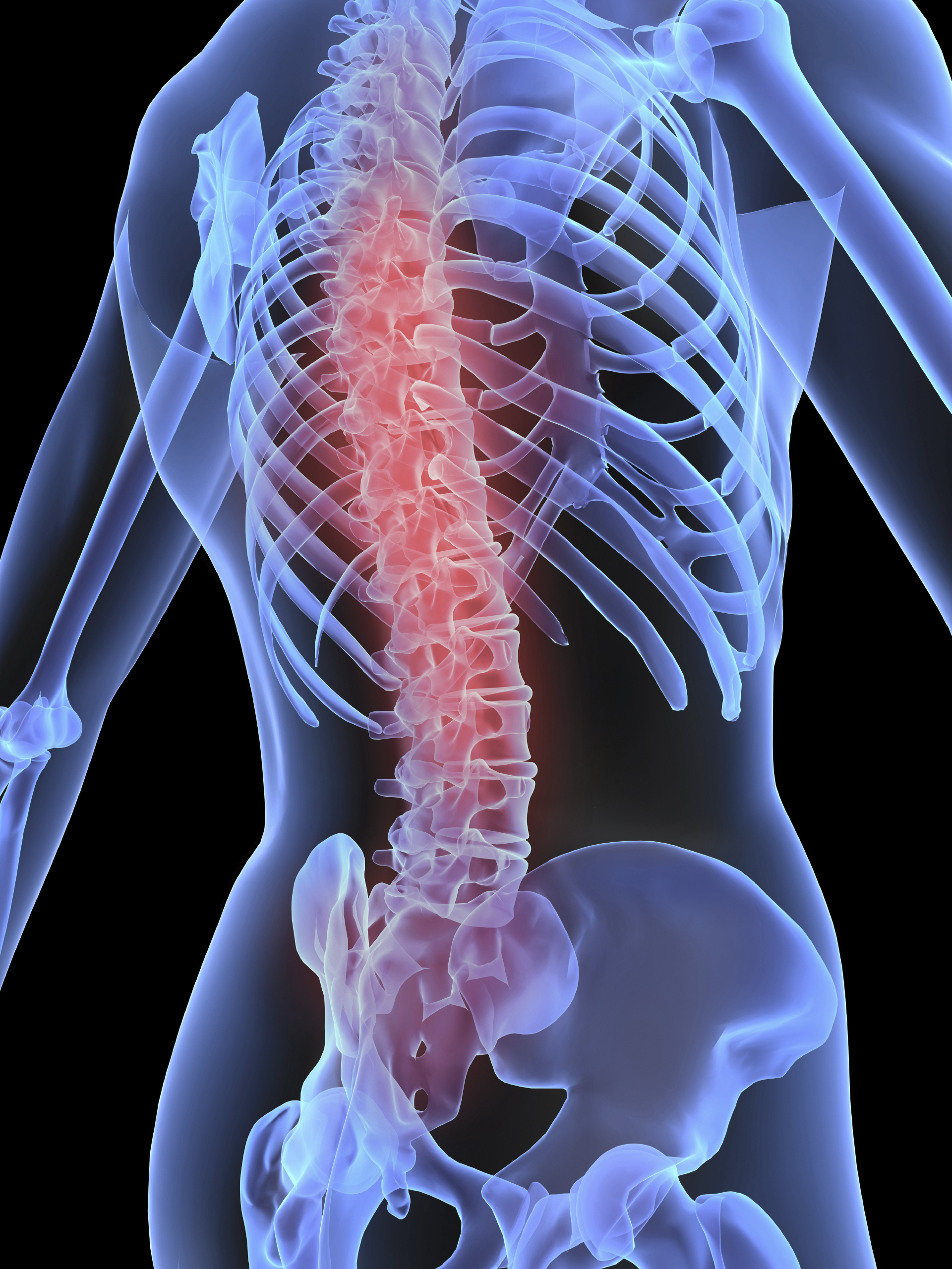
- Spinal cord injuries often result in damage to sensory nerve fibers that carry signals from the body to the brain, this can result in impaired bladder, bowel, and sexual function. Researchers are now investigating how damaged sensory nerve fibers can be repaired.
- Studies conducted using animal models, have identified three molecules that are associated with neural regeneration (α9 integrin, kindlin-1 and chondroitinase), specifically for the benefit of upper limb function.
- Researchers now hope to deliver these molecules to additional nerves in the hopes of restoring bladder function. To do this, researchers will investigate the most effective methods for delivering these molecules at a biochemical level. They will then determine which nerves are best suited to receiving these injections.
In a nutshell
Sensory fibers are specialized nerve fibers responsible for transmitting sensory information from the body to the brain. This allows us to recognize certain sensations such as: the need for a bowel movement, bladder fullness, or sexual arousal. Damage to sensory fibers following spinal cord injury can affect various bodily functions, including those related to the bladder, bowel, and sexual activities.
Previous studies using animal models have demonstrated that injecting certain molecules can promote the regeneration of sensory fibers and improve sensory function in the upper limbs. Three molecules have been identified, the first two called α9 integrin and kindlin-1 have been shown to trigger a growth response in the fibers, whilst the third molecule called chondroitinase has shown that it can overcome scar tissue. Researchers will build on these findings to better understand how these same molecules can be delivered to other sensory nerves, responsible for bladder function.
How this supports our goal to cure paralysis.
The wall of the bladder is made up of a collection of muscle fibers which are known as the detrusor muscle. Muscle fibers are woven together in a way that allows the bladder to stretch and contract in response to the presence of urine.
To coordinate this activity, nerves send messages via the spinal cord to various muscles, including the detrusor muscle in the bladder. In individuals with spinal cord injury, these signals are disrupted due to damaged nerve fibers. Researchers hope that their work can allow for the regeneration of these nerve fibers to restore bladder function, ultimately allowing individuals to regain their dignity and independence.
You may also be interested in


